Robotic-Assisted Proximal Gastrectomy with a Laparoscopic-Assisted Double-Tract Reconstruction for Proximal Early Gastric Cancer
1Seoul National University Hospital
2Dana-Farber Cancer Institute
3Brigham and Women’s Hospital
Main Text
Table of Contents
At most institutions caring for patients with early gastric cancer (EGC), tumors arising in the upper third of the stomach are usually managed with total gastrectomy and Roux-en-Y esophagojejunostomy. Given the impaired quality of life related to associated reflux and vitamin deficiencies, several high-volume centers have sought alternative gastrectomy and reconstruction strategies to total gastrectomy. In this case, a patient with EGC in the cardia found on screening endoscopy undergoes robotic proximal gastrectomy with double-tract reconstruction. His postoperative course was unremarkable, and he was discharged on postoperative day 7. His pathology demonstrated no residual tumor after preoperative endoscopic submucosal dissection. This video demonstrates the technique of an experienced surgeon performing robotic proximal gastrectomy with double-tract reconstruction.
Early gastric cancer (EGC) located in the upper third of the stomach presents a surgical challenge due to proximity to the cardia. Traditionally, total gastrectomy (TG) has been the standard treatment for upper gastric cancer. However, recent data from Japan and South Korea suggest that proximal gastrectomy (PG) can be considered a viable alternative for upper third EGC.1, 2 Since upper third EGC rarely metastasizes to the lower portions of the stomach,3 PG is considered oncologically appropriate in this scenario.
The method of reconstruction following resection of upper third EGC remains a point of controversy. The traditional approach of esophagogastrostomy has been associated with a high rate of reflux esophagitis and anastomotic stricture, leading to a preference for TG.4 However, double-tract reconstruction (DTR) has emerged as a technique that may mitigate the risk of reflux. Three anastomoses are involved with DTR: esophagojejunostomy (EJ), gastrojejunostomy (GJ) located 10–15 cm below the EJ, and jejunojejunostomy (JJ) situated 20 cm below the GJ.5, 6 This technique is called "double-tract" because it provides two passages for food: one directly into the jejunum and another through the remnant stomach and duodenum before meeting up in the jejunum. Gastric emptying studies have reported approximately 40–50% of food enters the remnant stomach.7 This approach preserves the function of the remnant stomach, making it an attractive option to preserve absorptive function for patients at risk of anemia and vitamin B12 deficiency.
A recent prospective randomized controlled trial (KLASS-05) conducted by multiple centers in South Korea reported promising results for laparoscopic PG with DTR (LPG DTR) compared to laparoscopic TG (LTG). The study found that LPG DTR required less B12 supplementation, was associated with better quality of life scores (physical function and social functioning), and had comparable overall complication and reflux rates compared to LTG. Moreover, the two-year overall survival and disease-free survival were similar between the two groups.7 These findings support LPG DTR as a safe and function-preserving alternative to LTG for EGC located at the upper third of the stomach.
The patient in this case is a 71-year-old male with ASA 2 and a BMI of 25.4. He was initially diagnosed with high-grade dysplasia in the upper third of the stomach during a screening esophagogastroduodenoscopy (EGD). Per Korean national guidelines, screening EGD begins at 40 years old and continues every two years.1 However, subsequent examination after endoscopic submucosal dissection (ESD) revealed that the lesion was EGC with submucosal invasion and deep margin involvement. Given these findings, the patient was referred for surgical management.
The tumor was located along the posterior wall of the cardia in the upper third of the stomach. According to Korean guidelines, PG is indicated for EGC located in this region. The patient had several comorbidities, including hypertension, diabetes, and hyperlipidemia, for which he was taking medication. He was also on 81 mg of Aspirin, which was held for five days before the surgery. There was no past surgical history.
The patient did not exhibit any specific symptoms related to his diagnosis of EGC, which is common in cases of early detection from screening EGD. No notable physical exam findings were present.
A computed tomography (CT) scan with stomach protocol failed to visualize the gastric lesion, and a follow-up EGD revealed a scar at the ESD location. His imaging revealed an accessory left hepatic artery arising from the left gastric artery. Plain films, including abdomen and chest X-rays, did not reveal any abnormalities.
The surgical pathology report for this patient revealed no residual tumor and no lymph node metastasis. The final pathological stage was pT1bN0, corresponding to Stage IA, which does not indicate adjuvant therapy. The patient is currently undergoing surveillance every six months, alternating between EGD and CT scans, to monitor for recurrence.
In cases where ESD reveals EGC with submucosal invasion and deep margin involvement (non-curative ESD), Korean guidelines strongly recommend additional surgical management due to the risk of lymph node metastasis ranging between 2–20%.8–18 To address this risk, gastrectomy with lymphadenectomy should be performed.
Another emerging approach is the double flap technique (DFT) for reconstruction after PG. Some reports suggest that DFT may be superior to TG to reduce reflux esophagitis, improve nutritional status, and minimize postoperative morbidity.19, 20 However, it is notable that minimally invasive PG with DFT reconstruction requires complex suture techniques resulting in longer operative time, necessitating further investigation.
The primary goals of treatment for this patient with EGC in the upper third of the stomach are complete removal of the lesion, minimization of the risk of lymph node metastasis, and preservation of gastric function and overall quality of life. In this case, the patient underwent PG with DTR to preserve the function of the remnant stomach and address the submucosal invasion necessitating re-excision of the involved deep margin following non-curative ESD.
Patients with EGC in the upper third of the stomach can benefit from PG with DTR by preserving absorptive and digestive gastric functions while effectively treating the malignancy. This approach is suitable for patients that meet criteria for EGC with favorable surgical risk profiles. Contraindications for this procedure include patients with advanced stage gastric cancer beyond the criteria for EGC (T1) or those who are not suitable for surgery due to severe comorbidities or other contraindications. The choice of treatment should be made on a case-by-case basis after careful evaluation of the patient condition and individual factors.
With the patient supine and left arm tucked in the adducted position, the abdomen is entered using the Hasson technique through a supraumbilical incision. Four robotic trocars are placed 8–10 cm apart above the level of the umbilicus, a 12-mm assistant port is placed near the right midclavicular line between the left arm port and the camera port, and the robot is docked. The surgeon moves to the robotic console, and the assistant remains at bedside to aid in retraction. The hepatogastric ligament is entered seeking any anomalous hepatic artery anatomy. If an accessory left hepatic artery is encountered, this is usually divided given its proximity to the tumor in the upper third of the stomach. If possible, a replaced left hepatic artery should be preserved.
The lesser sac is entered beginning at the bare area of the gastrocolic ligament. The greater curve is mobilized up to the left crus dividing the short gastric and left gastroepiploic vessels along the way. The omentum is split along a sagittal plane to permit tensionless positioning of the multiple anastomoses to follow.
Beginning at the midpoint between the right and left gastric arteries, nodal tissue is swept to the left along the lesser curvature to dissect station 3a nodes proximal from the left gastric artery into the specimen while preserving station 3b nodes and the right gastric artery. The stomach is divided at this landmark with 60-mm purple loads on an Endo GIA stapler. Station 8a nodes anterior to the common hepatic artery are dissected out and swept to the left along the lesser curvature into the specimen. The left gastric artery and vein are divided.
The abdominal esophagus is circumferentially skeletonized taking the vagus nerves in the process. At this point, the surgeon returns to the bedside to place a clamp on the specimen side of the esophagus and deploys a laparoscopic purse-string suture device proximally. Hook electrocautery is used to divide the esophagus in between freeing the specimen for retrieval in a specimen bag. An anvil for the 25-mm EEA stapler is secured into the esophagus with the purse-string. The robotic port between the camera at the umbilicus and the furthest right arm robotic port is converted to a 5-cm transverse port to remove the specimen and place an Alexis retractor to maintain insufflation for the latter portion of the case. The gastrectomy specimen is opened to send the proximal and distal margins of the stomach as well as lymph nodes by basin to pathology for review.
Given the position of the tumor is generally in the cardia or high body to permit proximal gastrectomy, lymph node basins 1, 2, 3a, 4, 7, 8a, and 9 are retrieved through a D1+ lymphadenectomy.
The three anastomoses are performed in retrograde fashion. The ligament of Treitz is first identified, and the jejunum is exteriorized where it is divided 15 cm distal from that point with a 60-mm purple load on the electronic Endo GIA stapler. From the distal cut edge of the jejunum or Roux limb, incisions are made in the jejunum 15 cm and 35 cm distally for the gastrojejunostomy and the jejunojejunostomy, respectively.
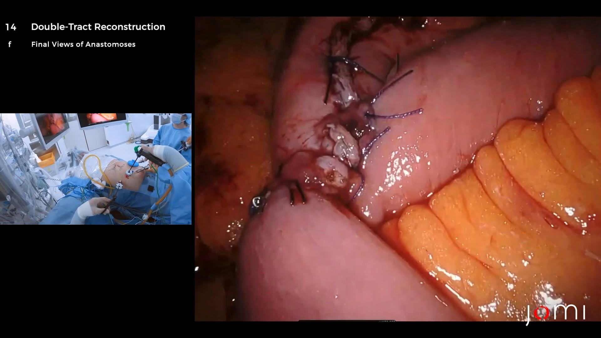
An extracorporeal jejunojejunostomy is performed first in stapled, side-to-side fashion with the 60-mm purple load on the electronic Endo GIA stapler, and the common channel is oversewn. An extracorporeal gastrojejunostomy is next performed in a stapled, end-to-side fashion 20 cm proximal from the jejunojejunostomy, and the common channel is oversewn. Finally, the staple line on Roux limb is removed and a 25-mm EEA stapler is inserted through this enterotomy with a sterile glove along the shaft to attach on the Alexis retractor to maintain insufflation. An intracorporeal esophagojejunostomy is then performed in a stapled, end-to-side fashion 15 cm proximal from the gastrojejunostomy as the spike from the EEA stapler is advanced out of the antimesenteric border of the jejunum before it is married to the anvil in the esophagus. The proximal esophageal donut is sent for final margin assessment. The overhang on the jejunum through which the stapler was inserted is excised with a single firing of the 60-mm purple load on the electronic Endo GIA stapler. The gastrojejunostomy is secured to the mesentery to minimize mobility. A nasogastric tube is inserted just beyond the esophagojejunostomy, and a drain is placed posterior to the gastrojejunostomy and esophagojejunostomy.
At experienced centers, PG with DTR can be performed with minimal morbidity and mortality. The recent KLASS-05 multi-center randomized trial comparing this technique to LTG found no difference in 2-year morbidity or overall survival; however, patients that underwent LPG with DTR needed less vitamin B12 supplementation and had improved physical and social functioning quality scores.7 Another reconstruction option after PG includes double flap reconstruction (DFR) where an anti-reflux valve is fashioned at the esophagogastric anastomosis.21 Most centers performing DFR at high volume do not additionally perform DTR; therefore, level I data does not currently exist comparing these techniques.
Following robotic PG with DTR, this patient had an unremarkable recovery. On the third day after surgery, the patient initiated sipping water. Between the fourth and fifth postoperative days, they transitioned to a fluid diet. They were advanced to full diet by postoperative day 7 when they were discharged home without the drain.
Final Pathologic Diagnosis
Proximal gastrectomy: no residual tumor, post-endoscopic resection status, tubular adenocarcinoma, well differentiated, confined to the submucosa (see synoptic report). There is no evidence of malignancy in 28 lymph nodes (0/28).
Synoptic Report
Tumor stage Summary: pT1bN0.
Tumor size (greatest dimension): 2.4x1.3 cm (in ESD slide).
WHO classification: tubular adenocarcinoma, well differentiated.
Depth of invasion: submucosa (in ESD slide).
Depth of submucosal invasion: 710 um (in ESD slide).
Width of submucosal invasion: 4.9 mm (in ESD slide).
Primary tumor: pT1b (tumor invades submucosa).
Small vessel (blood/lymphatic) Invasion: absent.
Large vessel (venous) invasion: absent.
Perineural invasion: absent.
Proximal esophageal margin: not involved by invasive carcinoma.
Distal stomach margin: not involved by invasive carcinoma.
Regional lymph nodes: pN0 (no regional lymph node metastasis): number of lymph nodes examined: 28 (LN#1, 0/8; LN#2, 0/1; LN#3a, 0/6; LN#3b, 0/0; LN#4d, 0/0; LN#4sa, 0/2; LN#4sb, 0/0; LN#7, 0/2; LN#8, 0/3; LN#9, 0/6; LN#11p, 0/0).
The Da Vinci Xi robotic platform is used for this operation with the Cadiere, vessel sealer, and tip up instruments. The Harmonic is used for extracorporeal division of the jejunal mesentery to maintain efficient hemostasis during dissection. The stomach and jejunum are divided with purple loads on the electronic Endo GIA stapler with tri-staple technology (0.95–1.12-mm staple height). The esophagojejunostomy is performed with a 25-mm EEA stapler (4.8-mm staple height) through an Alexis retractor. A novel laparoscopic purse-string device developed by colleagues at our institution is employed to assist with esophageal transection and suture fixation around the EEA stapler anvil.
Nothing to disclose.
The patient referred to in this video article has given their informed consent to be filmed and is aware that information and images will be published online.
Citations
- Kim TH, Kim IH, Kang SJ, et al. Korean practice guidelines for gastric cancer 2022: an evidence-based, multidisciplinary approach. J Gastric Cancer. 2023;23(1):3-106. doi:10.5230/jgc.2023.23.e11.
- Japanese Gastric Cancer Association. Japanese Gastric Cancer Treatment Guidelines 2021 (6th edition). Gastric Cancer. 2023 Jan;26(1):1-25. doi:10.1007/s10120-022-01331-8.
- Lee S, Son WJ, Roh YH, et al. Indication of proximal gastrectomy for advanced proximal gastric cancer based on lymph node metastasis at the distal part of the stomach. Ann Surg Open. 2021;2(4):e107. doi:10.1097/AS9.0000000000000107.
- Nakamura M, Yamaue H. Reconstruction after proximal gastrectomy for gastric cancer in the upper third of the stomach: a review of the literature published from 2000 to 2014. Surg Today. 2016;46(5):517-527. doi:10.1007/s00595-015-1185-4.
- Ahn SH, Jung DH, Son SY, Lee CM, Park DJ, Kim HH. Laparoscopic double-tract proximal gastrectomy for proximal early gastric cancer. Gastric Cancer. 2014;17(3):562-570. doi:10.1007/s10120-013-0303-5.
- Jung DH, Ahn SH, Park DJ, Kim HH. Proximal gastrectomy for gastric cancer. J Gastric Cancer. 2015;15(2):77-86. doi:10.5230/jgc.2015.15.2.77.
- Park DJ, Han SU, Hyung WJ, et al. Effect of laparoscopic proximal gastrectomy with double-tract reconstruction vs total gastrectomy on hemoglobin level and vitamin B12 supplementation in upper-third early gastric cancer: a randomized clinical trial. JAMA Netw Open. 2023;6(2):e2256004. doi:10.1001/jamanetworkopen.2022.56004.
- Kim ER, Lee H, Min BH, et al. Effect of rescue surgery after non-curative endoscopic resection of early gastric cancer. Br J Surg. 2015;102(11):1394-1401. doi:10.1002/bjs.9873.
- Choi JY, Jeon SW, Cho KB, et al. Non-curative endoscopic resection does not always lead to grave outcomes in submucosal invasive early gastric cancer. Surg Endosc. 2015;29(7):1842-1849. doi:10.1007/s00464-014-3874-2.
- Hatta W, Gotoda T, Oyama T, et al. Is radical surgery necessary in all patients who do not meet the curative criteria for endoscopic submucosal dissection in early gastric cancer? A multi-center retrospective study in Japan. J Gastroenterol. 2017;52(2):175-184. doi:10.1007/s00535-016-1210-4.
- Kawata N, Kakushima N, Takizawa K, et al. Risk factors for lymph node metastasis and long-term outcomes of patients with early gastric cancer after non-curative endoscopic submucosal dissection. Surg Endosc. 2017;31(4):1607-1616. doi:10.1007/s00464-016-5148-7.
- Kikuchi S, Kuroda S, Nishizaki M, et al. Management of early gastric cancer that meet the indication for radical lymph node dissection following endoscopic resection: a retrospective cohort analysis. BMC Surg. 2017;17(1). doi:10.1186/s12893-017-0268-0.
- Sumiyoshi T, Kondo H, Fujii R, et al. Short- and long-term outcomes of endoscopic submucosal dissection for early gastric cancer in elderly patients aged 75 years and older. Gastric Cancer. 2017;20(3):489-495. doi:10.1007/s10120-016-0628-y.
- Suzuki S, Gotoda T, Hatta W, et al. Survival benefit of additional surgery after non-curative endoscopic submucosal dissection for early gastric cancer: a propensity score matching analysis. Ann Surg Oncol. 2017;24(11):3353-3360. doi:10.1245/s10434-017-6039-4.
- Toya Y, Endo M, Nakamura S, et al. Clinical outcomes of non-curative endoscopic submucosal dissection with negative resected margins for gastric cancer. Gastrointest Endosc. 2017;85(6):1218-1224. doi:10.1016/j.gie.2016.11.018.
- Jeon MY, Park JC, Hahn KY, Shin SK, Lee SK, Lee YC. Long-term outcomes after noncurative endoscopic resection of early gastric cancer: the optimal time for additional endoscopic treatment. Gastrointest Endosc. 2018;87(4):1003-1013.e2. doi:10.1016/j.gie.2017.10.004.
- Kim HJ, Kim SG, Kim J, et al. Clinical outcomes of early gastric cancer with non-curative resection after pathological evaluation based on the expanded criteria. PLoS One. 2019;14(10):e0224614. doi:10.1371/journal.pone.0224614.
- Iwai N, Dohi O, Naito Y, et al. High-risk comorbidity influences prognosis in early gastric cancer after noncurative endoscopic submucosal dissection: a retrospective study. Dig Dis. 2021;39(2):96-105. doi:10.1159/000510115.
- Hayami M, Hiki N, Nunobe S, et al. Clinical outcomes and evaluation of laparoscopic proximal gastrectomy with double-flap technique for early gastric cancer in the upper third of the stomach. Ann Surg Oncol. 2017;24(6):1635-1642. doi:10.1245/s10434-017-5782-x.
- Shoji Y, Nunobe S, Ida S, et al. Surgical outcomes and risk assessment for anastomotic complications after laparoscopic proximal gastrectomy with double-flap technique for upper-third gastric cancer. Gastric Cancer. 2019;22(5):1036-1043. doi:10.1007/s10120-019-00940-0.
- Kinami S, Nakamura N, Tomita Y, et al. Precision surgical approach with lymph-node dissection in early gastric cancer. World J Gastroenterol. 2019;25(14):1640-1652. doi:10.3748/wjg.v25.i14.1640.
Cite this article
Narayan RR, Kim JC, Park DJ. Robotic-assisted proximal gastrectomy with a laparoscopic-assisted double-tract reconstruction for proximal early gastric cancer. J Med Insight. 2024;2024(427). doi:10.24296/jomi/427.