Pancreatic Debridement via Sinus Tract Endoscopy
15499 views
Procedure Outline
Table of Contents
- 1. Introduction
- 2. Surgical Approach
- 3. Exchange Superior Drain for 30 French Sheath
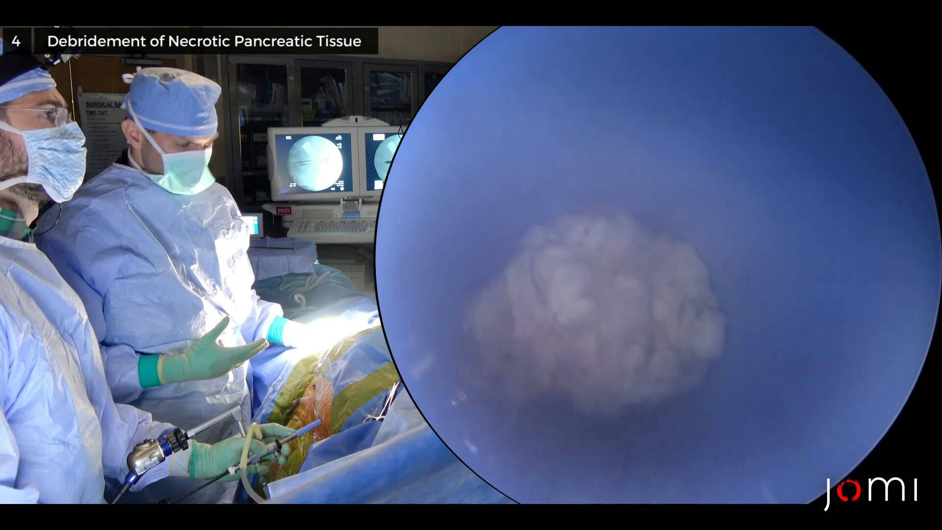
- 4. Debridement of Necrotic Pancreatic Tissue
- 5. Remove Sheath and Replace Drain
- 6. Suture Drain
- 7. Drain Study to Examine Fistula
- 8. Exchange Inferior Drain for 30 French Sheath
- 9. Examination of Cavity for Necrotic Tissue
- 10. Remove Sheath and Replace Drain
- 11. Drain Study
- 12. Suture Drain
- 13. Post-op Remarks
- Patient Positioning: A partial lateral decubitus position is often required to allow the percutaneous drain to be prepped into the field.
- Patient Preparation: Use a draping system that can catch the continuous irrigation. We use a neurosurgical head drape with attached collection bag.
- Set up and align fluoroscopy.
- Insert Super Stiff Amplatz guidewire into drain.
- Remove drain over the wire. Use fluoroscopy to be sure the wire stays in position.
- Enlarge incision to 1 cm.
- Insert dilating balloon over guidewire and inflate.
- Advance 30 French sheath over balloon.
- Deflate and remove balloon, keeping the wire in place.
- Insert the nephroscope to confirm placement in necrotic cavity, and remove the guidewire.
- Take care to avoid viable tissue on cavity borders.
- Remove entire scope and grasper—do not try to pull necrosis through the scope.
- Can work with or without continuous irrigation. Use whichever technique provides better visibility.
- Optional depending on cavity.
- Same as step 3.
- Same as step 4.
- Same as step 5.
- Same as step 7.
- Same as step 6.