Transmastoid Repair of Superior Semicircular Canal Dehiscence
Transcription
CHAPTER 1
All right, this patient - we're doing a left superior - transmastoid superior canal dehiscence repair, and he has a confirmed superior canal dehiscence on this side with symptoms of autophony, ear fullness, which is actually his major complaint, and dizziness. So, this is how we repair it. So, 15 blade. And we get down into the layer of the temporal parietal fascia down below it - through that into the loose areolar tissue, which gives us next bloodless plane. Get through the fascia. There we go - pops open. Okay, getting closer to the ear canal. Lift this up a little bit more here, and as you get closer to the ear canal, you start to see some blood vessels running right in front of the ear canal. It tells you you're about as close as you need to get. And that's right about here. So that's as far - close as we need to get there, and now we have it exposed. Can you hold the ear, please? You have a bipolar? Dry it up a little bit. And get all this dry so it doesn't bleed on us during the case. Okay. Can I have the bed towards me a little bit, please? I'll take the ear. All right, I'll take a Bovie. So, now we'll make our T-incision. And that gets up there close to the - hold ear, please - and I'll take a bipolar - to the deep temporal artery. And we have suction? And bipolar cautery usually works better up here than monopolar, so you can really grab the tissue and get both ends of the artery. All right, and I'll take the Bovie back. I'll take the ear. Okay, hold the ear, and I'll take a Lempert. Bovie the periosteum. Take that. Okay. Up to the ear canal, right there. This is as close as we need to get, and I'll take a Weity. So this gives us - all the way up to - suction - all the way up to the zygomatic root, so we can get really anterior - so we have enough room. All right, Bovie. And I'll just remove these attachments off of the - of the sternocleidomastoid - off of the mastoid tip. These don't come off very easily with the Lempert elevator. They're pretty adherent, so I always Bovie these off of - get the whole mastoid exposed. Now I have ear canal, tegmen, mastoid tip - got the whole thing exposed. So now we can switch to the microscope.
CHAPTER 2
All right, so I'll start with 5 cutter. So the thing to be aware of with this is the tegmen is often very thin and with multiple - multiple dehiscences, so you got to be really careful when you're drilling not to injure the dura. All right, water on. We'll start at the linea - linea temporalis, which is a good approximation for where the middle fossa is. Make sure you've got air cells there. See how that drill can get stuck in little holes and skip. So you always want to - you never want to plunge, and if there are holes, you always want to smooth them out. All right, so now I'm going to come along the ear canal, and then take the cortex off. So that's pretty much the cortical mastoidectomy, and let me have irrigation. It's real important to keep the bone dust off the field and keep it really clean, because it can dull the blade, it can dull the burr, and it'll also obscure your view. So... Okay, water down just a hair. Up. Very small difference between too little and too much. All right, so, now what we do is we follow the tegmen and making room, All these air cells need to go. So there we see the first glimpse of the tegmen, right through there. You just hold here. Gimmick. So we see as we lose the air cells, this is a very thin bone, and that's - we're seeing the the dura. You get a little better focus. We're seeing all the epidural blood vessels right here through this very thin bone here. That's what you go to be real careful of. Drill. So the idea is you find the tegmen, and then you catch up inferiorly and posteriorly. So I move deeper here and then open it up superiorly and remove all these air cells. You can see all the little - in a well-aerated mastoids, these little bone chips break off very - pretty easily. All right, so let me have an irrigation again. All right, drill. So I'm just opening it up widely posteriorly and inferiorly, making sure it's nice and saucerized. And by saucerized, I mean it's widest at the tip - out on the outside. Bipolar. So no overhanging ledges. Drill. Good. Saucerizing the ear canal there - back to the ear canal, and we're starting to get into the antrum. And now we're exposing the antrum. You see the lateral canal pretty nicely there, and I'm going to change my angle, so I can look into the - this direction more, and we're going to start seeing the incus. By pinning the ear canal, I can now - get closer into the antrum. We don't need to do much posteriorly there. So we do need to get all the way into the antrum. So it's always good to use as big a drill bit as you can. Let me have a 4 cutter - a 4 diamond, I mean. Water off. And a Gimmick. So I'm just starting to see the incus here. There's the incus. And this is the lateral semicircular canal. So this gives me my landmarks for how I'm going to be able to find the other canals. So here we have - this isn't actually an exposed area of dura. There's a natural dehiscence in the bone there, which can happen, so you just want to be real careful of the dura there. Don't want to injure it. So I'm going to polish this up. A little water on. Get some of the bleeding. The diamond is very good for polishing bone. It doesn't tear dura. Water off. Here's another area of exposed natural dehiscence right there. Let me see the bipolar. So these are just things you have to be super careful of, because these people have very thin tegmen, and if they didn't have a thin tegmen, they wouldn't have superior canal dehiscence. So it just kind of goes with the territory. Can I have bone wax on the back of a Freer? Which reminds me, I want to collect... What I want to do now is get some bone pâté. Can you put a 4 cutter on? I want to use some bone pâté to - to help plug the hole that I'm going to make in the lateral canal. 4 cutter. So I'm going to take some of this cortex. So I'm using not much water here - just keep it dry. All right, now I'll take a Freer. So I'll collect all this stuff. May I have a Petri dish? Dry. And I'll just collect this. And I'm going to mix this with bone dust - with - this bone dust with some bone wax to make a little paste that'll be really good for plugging the tiny little holes I'm going to make. And that should be plenty. All right, let me have irrigation. All right.
CHAPTER 3
So, now what I need to do is skeletonize the labyrinth a little bit more so I can get - start looking for my superior canal. And I know that my lateral canal is right here, so the superior canal is going to be coming above it - and posterior canal is going to be running 90 degrees here. Water on. And that tells me that I know the posterior canal and the superior canal will join together to form the common crus. Getting a little more working room here. Great, can I have a 3 cutter? So I'm starting to see here through this bone - water off. Let me have a Gimmick. So again, the lateral canal - so the posterior canal is going to be running orthogonal, 90 degrees to it, so right here's where the common crus is going to be. Superior canal is going to run like this. So when I clear these air cells out, I'm going to see the - the limbs of the posterior canal and superior canal coming together. Water on. All right, let me have a 3 diamond. Water off. We're getting near some dura. Don't want to mess with that. Water on. All right, water off. Let me see a Freer. Make sure we're in good focus here. All right, why don't you put a 2 diamond on. So what I'm looking for is a lot - water on. So right here - this is going to be posterior crus, and this is going - this is going to be superior canal, and this going to be posterior canal. This is where they're going to form the common crus. So superior canal is going to be going like this. And so what I'm going to want to do is make a hole in the anterior limb and a hole in the posterior limb here, and plug those with bone wax to keep - water off. Bipolar. So before I start doing that, I'm going to want to check the ABR. Make sure we're doing okay with that. So - and ABR is still real good, so what I'll do is make the bone wax pâté. So let me - let me see that - that stuff. Here we have our bone pâté. Let me have a ball of wax. Just a big ball. Get it kind of soft. And then kind of mix it all together here into one thing. You just - we'll just leave it up like that for now.
CHAPTER 4
And so, we'll start with the superior canal - the superior limb - or the anterior-most limb. Let me put on a 1 diamond. Well, I'll start with a 2, and I'll go in the back here. Okay.
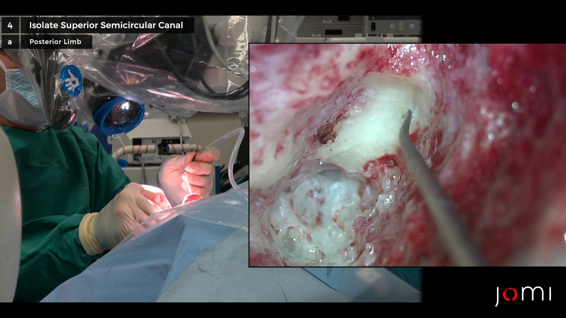
So, the limbs are going to be coming like this together, so this will be the posterior canal, and this will be superior canal. So I'm going to start looking right in here, and I'll just go real carefully. Water on. Just back-and-forth real carefully until I start seeing a blue-line. Zoom in just a little bit more. Can I get the bed away, please? All right. So I think I'm starting to get a sense right here. All right, let me have a 1 diamond. Drill carefully - a layer at a time. And... there we go. Water off. Right there's where I see it. Can you guys - does that show up? Yeah. All right, so I'm going to make that just a little bigger. Water on. All right, good. Water off. Let me have a Gimmick. So it's st- that's it right there. All right, so I'll check the ABR again. And the ABR is running. Do we have neuro-patties? How are we doing Holly? ABR is good. ABR is good, okay. So, let me have the drill one more time. I'm just going to real carefully thin eggshell this just a little more. Water on. All right, water off. Good. Now let me have the - put the blue suction on - blue suction tip. And there's a - and let me have a Rosen. So here we have a small hole. All right, let me have - let me have a dissector. Or a Gimmick. A Gimmick will work. And let me see the paste. Okay, now I'm going to put this over the hole. And let me have a bipolar and a neuro-pattie. And use that to pack it in. Not quite so wet. Can I have a - clean that? So we try to avoid suctioning over the open hole, but then we have it nice and plugged. Can I have another neuro-pattie? This shouldn't be drip - yeah, there we go. And we'll just - mash that in. All right, so that - Gimmick. So that was the hole right there, and now it's plugged. So that puts that all in there. And we'll check the ABR one more time. ABR is good.
All right, so now, dura - so you see how close it is. All right, why don't you put a smaller suction irrigator on? So the key is not to suction when you make the hole into the - into the canal, because that will cause deafness. All right, let's get the water on. All right, down. Down, down, down - keep going. Up - up a little. All right, I'll take a 1 diamond. And there's the blue-line, right there. Water off. Is that showing up okay? All right, let me have the blue suction and a Gimmick - I mean, a Rosen. All right. All right - oops - grab another little piece here. Good. All right, let me have a bipolar. And a neuro-pattie. And, push that in there. Let's check on ABR. ABR is still good. Excellent. Okay, so hearing's still good, and it looks like there might be just a tiny little bit of CSF coming out of there, so let me see a Freer. And let me have a 5 suction. Let me see the rest of the bone pâté. I'll pack this up here. And... call that good. And that's how you - that's how you do a transmastoid, two-holed approach for isolating a superior canal dehiscence.
CHAPTER 5
[No Dialogue.]
