Open Radical Cholecystectomy with Partial Hepatectomy for Gallbladder Cancer
Transcription
CHAPTER 1
I'm Hiromichi Ito, I'm head attending surgeon of hepatobiliary service at Cancer Institute Hospital in Tokyo, Japan. Cancer Institute Hospital is the oldest, private cancer institute in Japan, and in the country, we ranked number 1 as the highest volume center for the gastric cancer- colon cancer, and hepatobiliary cancer. As attending surgeon, I routinely doing to the many liver resections for the colorectal mets as well as primary liver cancer. And also, as a- as a common disease in Japan, we do have... biliary cancer and gallbladder cancer as today's case. Today we are going to see the case with gallbladder cancer. The patient is a 60-year-old gentleman who had bladder cancer 1.5 years ago, and it was treated by radical cystectomy with iliac conduit following the neoadjuvant chemotherapy. Fortunately, this disease was diagnosed at an early stage, and he has been following up with imaging, expectantly. And recently, he was pointed out as a- some gallbladder mass on the routine CT scan, and referred to the hepatobiliary service. So retrospectively, we are going back to the CT scan 9 months ago. There was a- they started to show a small nodules in the gallbladder wall, and over the 9-month period of time, it keeps growing. And certainly, the gallbladder cancer was suspected, and further work-up was done. Further work-ups include the EOB MRI to rule out the intrahepatic metastasis, and PET scan- most are negative for the distal metastasis. And- his tumor markers were CEA and CA 19-9 were both normal range, and therefore we recommend to the radical cholecystectomy including to the removal of the gallbladder, as well as the liver bed, and lymphadenectomy around the porta hepatis, and we can still show the case today. Okay, so the key steps of the procedure- so first we make the small midline incisions and explore for the peritoneal space to rule out- contraindication for the resections. We look for the peritoneal seedings, and we palpate the liver to look for- liver metastasis undetected on the perioperative imaging studies. And after we confirm there is no contraindications, the incision will be extended. I use- I prefer to use a reverse-L incision to provide the best exposure in the right upper quadrant organs. And we place on the liver retractor that maximizes the exposure around the liver, which was underlying the organs and the ribcage. After the incisions and retractors placed, we do the full exploration of the peritoneal space again. We have to carefully rule out for the peritoneal seedings, or liver metastasis. To rule out the liver metastasis, we perform the intraoperative ultrasound with IV contrast Sonazoid, and that can detect very small regions- less than 5 mm, and fortunately in this case, it was all negative. And then, we do the kocherizations, mobilize for the duodenum and the head of the pancreas, and expose to the IVC. This procedure was done for 2 reasons. The one is it provides us access to the literal pancreatic lymph node- number 14 lymph node stations. And also, it provides us the opportunity for the lymph node sampling in the para-aortic lymph node station. The para-aortic lymph node metastasis to the para-aortic lymph node stations is considered as N1 disease. Therefore, it's a contraindication for the definitive resections. So always we routinely sample the- lymph node, and send it to the frozen section. In case this lymph node proved positive for cancer, we abort the resection, and just close, and put the patient on systemic chemotherapy. In this case, we sent 1 big lymph node, and it was reported as negative for cancers, and we decided to go to the resection. And following that, we do the portal lymphadenectomy. It can be started with the excision of the retropancreatic lymph node or suprapancreatic lymph node, namely the common hepatic artery lymph node, number 8 lymph node station. We did start with the number 8 lymph node station dissections, and at that point, the key of this step is to retract to the pancreas gently towards to the leg, and that allows to the exposure of the common hepatic artery. In this case, the common hepatic artery was running very behind of the pancreas, and exposure was not easy, and we could not tape over these arteries. However, the lymph node above the common hepatic artery can be excised and completely removed. without taping to the common hepatic artery itself. And then we are doing the ligate and divide of the supraduodenal vessels, and expose to the common bile duct, and GDA, and hepatic arteries. Once the artery is identified and taped, the surrounding lymphatic tissues and connective tissues are excised sharply, and vessels and bile duct was skeletonized. And portal vein is underlying these structures. And covered by lots of lymph nodes, number 12 lymph node station, so that these lymph nodes are also excised, dissected off from the portal vein, and the portal vein is also circumferentially skeletonized and the taping is always helpful for this part of the procedure. And the dissection is from the lower to the higher levels, and continue. And once we encounter for the cystic duct, the cystic duct is isolated, and ligated, and divided. And at this point, we send- cystic duct margin was sent to the frozen section to confirm that the cancer is not invading to this area. If the- frozen section reported as positive for cancers, the operative plan was changed for the en-bloc resection for the common bile duct as well. Fortunately, in this case, the cystic duct margin was negative, so we can preserve for the entire length of the common bile duct. So the lymph node dissection is continued, and the left and right hepatic artery was completely skeletonized. The anatomy of the hepatic arteries need to be crystal clear on the perioperative CT scans because there is a lot of anomalies, and- if you don't know for the- arterial anatomy, like replacing the hepatic arteries behind- behind of the portal veins, you are injured of the structures that's causing to the disastrous consequences. And- once the right hepatic artery and the bile duct was dissected off to the liver hilum, then the attention was moved to the liver. The radical cholecystectomy includes the partial resection of segment IV and segment V as a part of the procedure, and you can remove the liver as a wedge resection or anatomical segmental resection. In this case, I do the segmental resection because there was a gross infiltration of the tumor into the liver, and I don't like to leave any positive margins there. But current researchers say there is no difference for the extended liver resection vs the wedge resection of the gallbladder fossa. For the liver resections, we do the Pringle maneuver, we clamp for the hepatic artery, separately, and portal vein clamped for 15 minutes, and 5 minutes released. The liver parenchyma was transected using the... clamp-crush technique. We crush the parenchyma with a clamp, which allows the underlying vessels exposed, and the small vessels can be ligated with a Ligasure, and Glissonean pedicles, including the bile duct, we prefer to ligate with a Vicryl suture. We divided segment IVB Glissonean pedicle first, and the dissection line down onto the hilar plate. The hilar plate was exposed widely, and that allowed for the identification of the Glissonean pedicles to the segment V. So the segment V is isolated and doubly ligated, and divided. Then you can see the demarcated lines between segment V and VI, which is the line for the right dissections. Dissection was continued from the right side and connected to the dissection plane, and then the specimen was taken. After the specimen was taken, we... the hemorrhage very carefully from the liver bed, and also the bile leak- as we expose to the hilar plate and bile duct, pretty wide range, it is a high risk for the bile leak as a- possible complication, so we do very carefully look at the bile leak, and if we found it, we can fix it with a 6-0 PDS sutures. In this case, fortunately there was no sign of a bile leak. And then we leave 2 drains, one is a Winslow, for the risk of the pancreatic leak because we removed the number 14 lymph node, and number 8 lymph nodes that sometimes torn the pancreatic capsule, so we always leave the drains in the Winslow- foramen of Winslow. And also, we leave the one drain in the liver resection surface. And we are not routinely place on the drains on the liver resection surface, but in this case, because of the wide exposure of the bile duct on the surface, we leave it. The peritoneal space was irrigated with 5 L of fluid, and then we closed routinely. Sometimes we do the gallbladder mass as suspect of the cancer, but this is- you know, a spectrum of disease, and some more looks like the benign disease, some is very clear malignancy. For the malignancy, we do- our approach is like this, we start with the completion of the lymphadenectomies, and we do the major segmental anatomical resections, and on the other hand, if the cancer is not so highly suspected, we do the cholecystectomy first with some small amount of the- the liver was included, and once we confirmed the diagnosis, we proceed for the rest of the procedure, namely the lymphadenectomy along the porta hepatis.
CHAPTER 2
So we start from the small incisions to decide resectability.
Cytology- peritoneal cytology, okay? So we took the- we put the catheter in the Douglas pouch, and we wash the- peritoneal fluid, and we send it to cytology to rule out the carcinomatosis. And also, I check the occult liver metastasis from the small incision, and we don't have one, then we extend the incision. Knife, please.
Okay. So we are taking the xiphoid process off to facilitate the exposure of the right upper quadrant.
Okay, go ahead, please. So we take the 1 cm xiphoid process, that significantly facilitates the exposure on this area, so we always- routinely remove the xiphoid process. Okay, let's take the round ligament.
So we take the round ligament as long as possible to use as a handle for the liver retractions, and always we ligate both sides of the round ligament because possible bleeding. Alright, let's move on to the side. So to divide the muscle, I use the Ligasure that facilitates the bloodless- incisions of the muscles. So we approximate the peritoneum to the muscles, that is- helping to- the good exposures. And also it is helpful to- when we close the fascia, identification will be much easier.
So we protect the wound with the towels, and then place on the retractors. Okay. So tumor is extending towards this length- it's pretty big.
CHAPTER 3
Well, actually we have some small nodule in the surface, could be the peritoneal seeding, we need to excise and biopsy before we commit to the resection. We can do that later.
Alright, meanwhile we can do the- ultrasound. Middle hepatic vein. So the tumor is here, extending down to this level, we can feel that hard nodule here as well- could be the lymph node. And exposing to the serosa. So most likely, it's more than T3. And because we do find for the- nodules in the liver surface, we have to rule out the metastasis. If it turns out to be metastasis, we need to abort the resections. It doesn't look like cancer, so we'll see how the frozen section say. Take a look at this.
So we explore the peritoneal space to rule out the carcinomatosis. So he has ileostomies, so we can't go too far. But I don't see any obvious nodules, so it might be okay. Okay. Alright, let's move on to the- Kocher's maneuver. And we can take down to the hepatic flexure. Okay, we can see the IVC now.
CHAPTER 4
So we have good news, cytology was negative for cancer. So this is the aorta. So the purpose of the Kocher's maneuver is to gain access to the retropancreatic lymph nodes as well as- we can do the- sampling of the para-aortic lymph nodes for the staging purpose. If there's a- para-aortic lymph node was positive for cancer, we may abort the definitive resections.
CHAPTER 5
So now, we start sampling of the number 16 para-aortic lymph node here. So the peritoneal nodule was negative for cancer, so I assume we are okay to proceed with the resections. Right angle. Thank you. Suction. Number 16 lymph nodes. So we completed the para-aortic lymph node sampling, and usually we wait for the frozen sections.
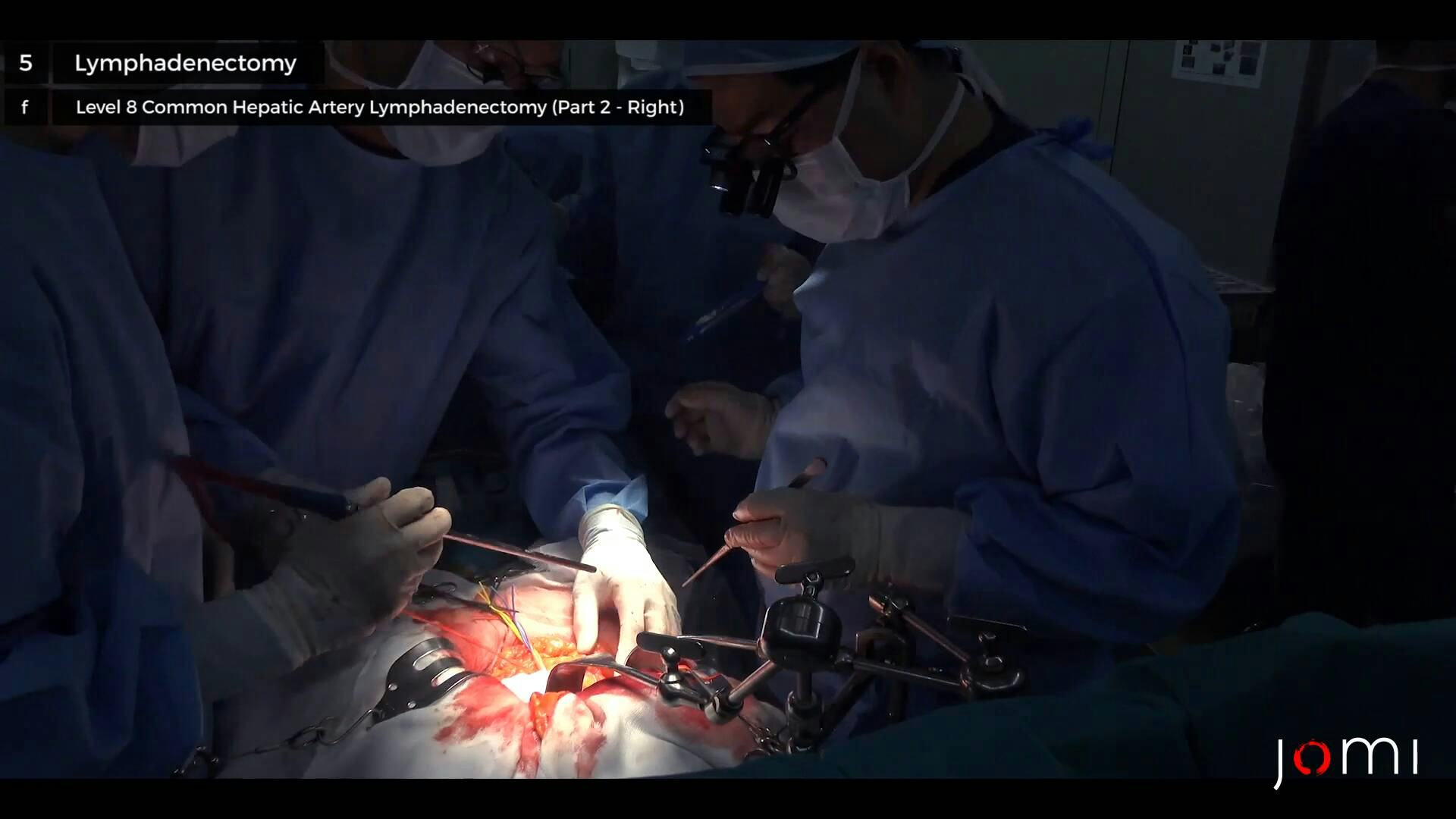
So we open the lesser sac. This is omentum. So we do retract the caudate process of the liver, that allows good exposure of the pancreas and- common hepatic artery and number 8 lymph node stations. So we are dissecting this part. So the assistant retracts the- pancreas, pull down. And we incise the peritoneum over the pancreas, which gains the space- access to the space between the lymph node and the common hepatic artery. So we think this is the right gastric artery. We can sacrifice later, but at this point, I- taped, and decide… Because the identification of the common hepatic turned out to be challenging, because of an enlarged lymph node, we approach for the right side of the porta hepatis, and find out the GDA, and then find for the common hepatic artery. So supraduodenal vessels- ligating and divided. Right angle. Okay, so the para-aortic lymph node was reported negative for cancer, so I think it's a green sign to proceed for the resection. We already had the middle of the lymphadenectomy, number 8 is relatively difficult because it's enlarged, and densely adhered- adherent to the common hepatic arteries, and we didn't see the common hepatic artery yet, completely. So I think the common hepatic artery is here. Take a look- this is the common. So let's go to the 13. So because number 8 lymph node is relatively… and bloody, so we started with the 13 first, and then we do- take care of the number 8 lymph node later. So number 13 lymph node is retropancreatic lymph node. So after the- the view of the extended Kocher maneuver, you can see the lymph node maybe around here to here- this area with the lymph node.
So we excise for the peritoneum, a little bit. And expose the surface of the pancreas. Very meticulous dissection is necessary. If you injure the pancreas, that- causing to the pancreatic leak, postoperatively. So we tape the common- common bile duct. And after this, we see the portal vein.
So we do the surface of the portal vein skeletonized now, and peel off all the lymphatic tissues and nerves- by sharp dissection. Right angle. So we ligate the small branch of the portal vein to avoid the bleeding. So we circumferentially dissected the portal vein, and we are taping to facilitate the retraction. Okay, so the right side, the portal- lymph node was dissected off from the portal vein and the bile duct, so we can connect the number 8 lymph node from the left side dissections, so we are going back to the number 8.
So we tape the left hepatic artery. And after that, we hope we can find the right hepatic artery. Right angle. So now we see the more anatomical structures. This is likely the right hepatic artery.
And this is the GDA. Right angle. Right angle. So we are almost done for the lymphadenectomy, and then we isolate the cystic duct. And confirm the negative margins- on the cystic duct margins.
Right angle. Right angle. So this is the cystic duct, and we divide the line here, in its root, and we confirmed of the cystic duct margin negative. If positive, we need to remove the entire common bile duct. So at this point, we divide it, and send bile duct margin in the frozen sections. We doubly ligate. So we divide the cystic duct.
So the anterior tissues was flipped out to the left side. Right angle. Right angle. Right angle. So now, we are dissecting the back side of the common bile duct. It's the right hepatic artery is running behind the common bile duct, and we dissected that space. Could be the cystic artery. Okay, so the number 18, number 12 is a- the lymph nodes are all en bloced and swiped towards the specimen, and only connection here, we can divide. Now it was reported- bile duct margin- cystic duct margin was negative for cancer, so that means we don't need to excise the common bile duct. Looks like the cystic duct is here. Right angle, please.
So this is the cystic arteries. Ligate it… Do one more- we do double ligate. Okay, and another 4, please. Okay, so the lymphadenectomy will be almost done, we're- once- artery and bile duct is- completely free from the cystic plate, we can move on to the hepatectomy.
Okay, so- I think that we are- completed the- hilar dissection, so- 5-0 Proline… So this is the completion of the lymphadenectomy.
We do see the common hepatic artery, right gastric arteries, and GDA, left and right hepatic artery, and the stump of the cystic artery. And the bile duct- extrahepatic bile duct is entirely skeletonized and likely that- left and right bifurcation is around here. And the portal vein is also coming behind, this is circumferencially- skeletonized. And number- 13 lymph node- retropancreatic lymph node was also removed en bloc. So this is the completion of the lymphadenectomy. And then we move on to the partial hepatectomy part.
CHAPTER 6
And this is all the lymph node dissected, and it's attached to the specimen side. Alright, let's move- before we starting to the hepatectomy, we mobilize to the right lobe of liver a little bit. That facilitates the- enhances the safety of the liver resection. So now we are mobilizing to the right lobe of liver that allows us to elevate the liver higher up that decreases the blood loss during the parenchymal dissection- transection. I think that's enough mobilization, so we are now to dissecting the- adrenal gland, and I think- just a little bit more. Okay, I think this is good enough.
So this is IVA and IVB, and- the pedicle IVB needs to be removed, and IVA needs to be preserved, so this is IVA. And we make the lines between the IVA and IVB. And for the tumor infiltrations, is- this levels. so that should be enough. And we can mark with a… And we don't know the exact demarcation line yet, but in the end, we- after we ligate the- the- segment V- Glissonean pedicles, we can see the demarcation line between the segments VI and V, most likely- somewhere here. This is just to estimate. So this line is diving onto the hilar plate, and we can connect on- just above the hilar plate, on this line. Right angle. So now, we start the- transecting the liver parenchyma.
So this is the pedicle IVA. So we- we'll have a 5-minute break, so 15 minutes for the- we clamp the in-flow, and we let the liver some bleeds- between, so every 15 minutes we open the clamp, and let the liver bleed. And then after 5 minutes, we declamp and start back again. So I think this is the middle hepatic vein branch. So this is one of the S5 branches. Okay, we ligate the segment V pedicle. So segment V should be ischemic now. Now, after you can see the very nice demarcations, it's almost exactly how I marked it before. Now we can divide the liver from the segment V side.
So this is the completion of the- extended cholecystectomy. This is the bile duct, completely skeletonized circumferencially. And right hepatic artery. Left hepatic artery and right gastric artery, branching off from the common hepatic artery running behind the pancreas. And all the soft tissues and lymph nodes are removed. And the portal vein is also circumferentially skeletonized. on the bottoms of the porta hepatis. And- for the liver resection side, segment IVB was removed, and the hilar plate is exposed. And this is the anterior Glissonean pedicles, and the posterior Glissonean pedicle is over here. And- segmental Glissonean pedicle V was ligated and divided at its root, so it's pretty much the anatomical segment V and IVB resections. And we do the- irrigate the abdomen, and place the drains, and close.
CHAPTER 7
Alright, so we are closing, and placing on the drains, and we place the drain after we close the transverse incision first. So we place the drains under the foramen of Winslow. So the frozen section examination confirms our diagnosis of the gallbladder cancer. He has poorly differentiated adenocarcinoma.
Thank you. Thank you very much.
CHAPTER 8
So, the case is done, and as you can see, the- we completed the- planned case as expected. And, however, on the first exploration, we found the tumor mass appear to be much bigger than expected, and grossly invading to the- to the liver, so I worry about for the peritoneal metastasis, but full exploration, including peritoneal washing cytology, were all negative, and only one nodule seen under the liver was excised, and proved negative for cancer. And also, we sampled the- routine staging, sample para-aortic lymph node as staging purpose, and it was proved as negative for cancer during the surgery, so I think it's a good indication for the- for the surgery. The surgery itself is going to be relatively smooth. unlike the- common Japanese operation, he's a little bit obese compared to the other type of- other patient with gallbladder cancers. So, lymph node dissections is a little bit more difficult than usual, but we completely skeletonized both hepatic arteries and the portal veins, and I don't believe that residual tumor left behind. So the- postoperatively, the patient was placed on the intensive care unit for the overnight observations, and most of the patients can return to the surgical floor on the post-day 1. Actually the possible course is going- pretty well- patient is doing pretty well after this big operation because we don't get touched with the bowel, and the patient can start the diet very smoothly. And drains was- we send the drain up to fluid every day, and check for the amylase and bilirubin levels, and on post-day 3, these levels within normal limits, we remove the drains on day 4 or day 3. And after that, the patient can eat the diet, and the pain is under control, the patient is ready to go home. Unlike the- insurance system in the United States, In Japan- insurance system- most of the patients tend to stay much much longer than the United States. But most of the patients are ready to go very short period of time. And regarding to the long-term outcomes for the gallbladder cancer, it depends on this patient's final stage. If the patient has lymph node metastases, that meaning to the stage 3 disease, the prognosis is very, very bad, and the likelihood of the recurrence is 70-80%, and we do recommend for the adjuvant treatment for this type of the patients. And on the other hand, if the lymph node was negative, stage 2 or stage- stage 1 or stage 2 disease, the outcome is pretty good- the 5-year survival in this hospital is greater than 75%, and most of the patients is doing well, and we don't recommend to adjuvant chemotherapy for this stage of the patient.