Closed Cephalomedullary Nailing of a Diaphyseal Femur Fracture on a Fracture Table
Main Text
Table of Contents
Midshaft femur fractures have an annual incidence of 10 per 100,000 person-years. Femoral fractures typically occur in two major settings: high-energy mechanisms related to trauma and low-energy mechanisms in insufficiency fractures observed in elderly patients with osteopenia. Patients present with pain, swelling, and limited range of motion. Intramedullary nailing is the definitive surgical treatment for femoral fractures to allow secondary healing of bone. Such a repair is performed here on a patient with a diaphyseal femoral fracture. Surgeon preference was to perform a closed cephalomedullary nailing with the patient supine on a radiolucent fracture table for traction.
Femoral shaft fractures typically occur in two situations: high-impact trauma and low-impact fractures in elderly patients with osteopenia. Definitive treatment is surgery with an intramedullary rod to allow secondary healing of bone.
The patient, in this case, is a 76-year-old woman with a history of osteoporosis and total knee arthroplasty (TKA) treated with bisphosphonates for over five years who presented with a diaphyseal femoral fracture that occurred while ambulating.
Femoral fractures present with pain and swelling of the affected thigh. On clinical exam, a restricted range of motion is observed. Shortening of the limb and gross deformity may be present. Patients should be evaluated to rule out concomitant neurovascular and soft tissue injury around the fracture site.
Anteroposterior (AP) and lateral radiographs of the femur are obtained to visualize the fracture line. Hip and knee radiographs are also obtained, which are important to rule out femoral neck fractures.1 Atypical femoral fractures are transverse and can be slightly oblique (<30 degrees). On AP view, there is often a “beak” visualized in the cortex.2 Although most atypical femoral fractures are in the subtrochanteric region or midshaft, this patient presented with a more distal fracture between the middle and distal third of the femoral shaft. In atypical femoral fractures, the contralateral limb should also be evaluated.
Midshaft femur fractures have an annual incidence of 10 per 100,000 person-years.3 While younger patients are more likely to present with midshaft femoral fractures in the setting of high energy trauma such as motor vehicle accidents, low-energy mechanisms, or spontaneous atraumatic mechanisms during activities of daily living are common in elderly patients.4 Both bone fragility due to osteoporosis and long-term bisphosphonate use have been linked to femoral fractures in elderly women.5 This patient had a low-energy mechanism of injury, as the fracture occurred while ambulating in the setting of a known medical history of osteoporosis and bisphosphonate use for over five years.
Femoral fractures are treated definitively with intramedullary fixation.6 Generally, retrograde nailing can be used. In contrast, osteoporosis-related fractures can be treated as pathologic fractures using antegrade nailing with an intramedullary rod via an interlocking nail. A more proximal fracture might require open reduction with the patient in a lateral position on the radiolucent fracture table. In this case of a closed fracture, a fracture table was used with the patient in the supine position to provide traction for the reduction.
The goals for treatment are reduction and fixation of the fracture to allow secondary healing of bone. Surgical techniques are designed to avoid rotational malalignment, nonunion, and destabilization of the prosthesis in this case.
Patients must be appropriate candidates for surgery in order to undergo definitive treatment of the fracture. External fixation can be performed immediately with intramedullary nailing in 2–3 weeks in cases of complicated trauma including severe open fractures and coexisting vascular injury.
Patients are monitored postoperatively for neurovascular injury, compartment syndrome, and infection.7, 8 Long-term complications can rarely include avascular necrosis, joint instability, and nonunion.6
Of paramount importance in obtaining adequate healing of the fracture is a look at the clinical management of patients such as the one seen here. Specifically, the need to immediately interrupt the antiresporptive medication (in this case, a bisphosphonate) and to observe the rate of bone healing, as many authors recommend the adoption of anabolic measures by administering teriparatide (r-PTH hormone).
In this case, a closed diaphyseal femur fracture was treated with cephalomedullary nailing on a fracture table for reduction. The patient is a 76-year-old woman with a history of osteoporosis, long-term bisphosphonate therapy, and a well-functioning prosthetic knee.
Bisphosphonate use over longer treatment courses may be linked to an increased incidence of femoral shaft fractures.4 However, the incidence of both typical and atypical fractures is higher for patients with osteoporosis who are not receiving bisphosphonate therapy.9 Because long-term bisphosphonate therapy is a risk factor for atypical fractures, it is unclear if patients continue to benefit from treatment for longer than five years in postmenopausal osteoporosis.10
Intramedullary nailing is the standard treatment for femoral shaft fractures and is associated with good outcomes.6 There is a low incidence of nonunion and other complications. Antegrade intramedullary nailing is the standard treatment for diaphyseal femur fractures with improvement in rates of malalignment.11, 12 The Trochanteric Fixation Nail (TFN) used in this case is positioned in order to avoid misalignment during fixation using the greater trochanter as a starting point.13 Antegrade nailing can also be performed using the piriformis as a starting point, though the injury to abductor muscles can be observed as a postoperative complication.14 Immediate postoperative complications to evaluate patients for include pudendal nerve palsy.7 There are cases of compartment syndrome associated with the use of a fracture table for traction, which can be avoided with careful positioning of the contralateral leg.8, 15
Nonunion is the most common cause of failure of the fixation of diaphyseal femoral fractures.16 Intramedullary nailing fixation has low risks of nonunion, with nonunion usually observed in patients with hypertension, obesity, and diabetes, or fractures in the proximal third junction.17 A retrospective analysis of 51 patients undergoing intramedullary nailing of femoral shaft fractures with third fragments showed that delayed union was affected by the displacement of the third fragment.18 Due to the altered bone biology in patients such as the one here, some adjuvant drug therapy may be needed to help reduce the risk of developing a nonunion.
Further research is indicated in related cases of atypical femoral fractures. The optimal treatment regimen of bisphosphonate therapy in postmenopausal osteoporosis continues to be an active area of research, with new data emerging on specific populations who benefit from the reduced risk of hip fracture.5 Systematic reviews often focus on identifying risk factors and radiographic evidence for nonunion in patients undergoing fixation to reduce reoperation rates.19 Robotic techniques are a newer area of research under investigation for alignment in intramedullary nailing for femoral fractures.20
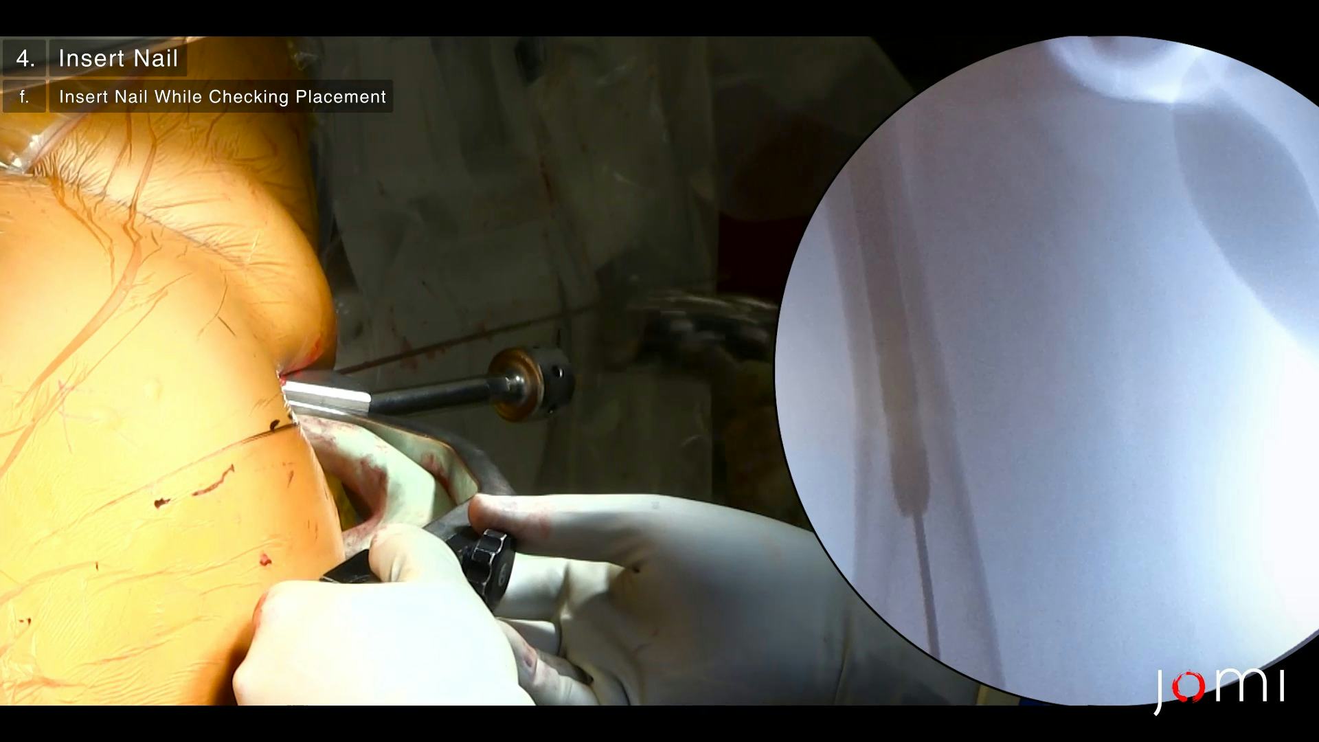
Cephalomedullary nailing system the DePuy Synthes Trochanteric Fixation Nail (TFN) was used in this case.
Nothing to disclose.
The patient referred to in this video article has given their informed consent to be filmed and is aware that information and images will be published online.
Citations
-
Tornetta III P, Kain MSH, Creevy WR. Diagnosis of femoral neck fractures in patients with a femoral shaft fracture: improvement with a standard protocol. JBJS. 2007;89(1):39-43.
https://doi.org/10.2106/jbjs.f.00297 - Marshall RA, Mandell JC, Weaver MJ, Ferrone M, Sodickson A, Khurana B. Imaging Features and Management of Stress, Atypical, and Pathologic Fractures. Radiographics. 2018;38(7):2173-2192. https://doi.org/10.1148/rg.2018180073
- Weiss RJ, Montgomery SM, Al Dabbagh Z, Jansson K-Å. National data of 6409 Swedish inpatients with femoral shaft fractures: stable incidence between 1998 and 2004. Injury. 2009;40(3):304-308. https://doi.org/10.1016/j.injury.2008.07.017
- Isaacs JD, Shidiak L, Harris IA, Szomor ZL. Femoral insufficiency fractures associated with prolonged bisphosphonate therapy. Clin Orthop Relat Res. 2010;468(12):3384-3392. https://doi.org/10.1007/s11999-010-1535-x
- Black DM, Geiger EJ, Eastell R, et al. Atypical Femur Fracture Risk versus Fragility Fracture Prevention with Bisphosphonates. N Engl J Med. 2020;383(8):743-753. https://doi.org/10.1056/NEJMoa1916525
- Ricci WM, Gallagher B, Haidukewych GJ. Intramedullary nailing of femoral shaft fractures: current concepts. J Am Acad Orthop Surg. 2009;17(5):296-305. https://doi.org/10.5435/00124635-200905000-00004
- Brumback, R. J., Ellison, T. S., Molligan, H., Molligan, D. J., Mahaffey, S., & Schmidhauser, C. (1992). Pudendal nerve palsy complicating intramedullary nailing of the femur. The Journal of Bone & Joint Surgery, 74(10), 1450–1455. https://doi.org/10.2106/00004623-199274100-00003
- Anglen J, Banovetz J. Compartment syndrome in the well leg resulting from fracture-table positioning. Clin Orthop Relat Res. 1994;301(301):239-242. https://doi.org/10.1097/00003086-199404000-00037
- Rizzoli R, Akesson K, Bouxsein M, et al. Subtrochanteric fractures after long-term treatment with bisphosphonates: a European Society on Clinical and Economic Aspects of Osteoporosis and Osteoarthritis, and International Osteoporosis Foundation Working Group Report. Osteoporos Int. 2011;22(2):373-390. https://doi.org/10.1007/s00198-010-1453-5
- Donnelly E, Saleh A, Unnanuntana A, Lane JM. Atypical femoral fractures: epidemiology, etiology, and patient management. Curr Opin Support Palliat Care. 2012;6(3):348-354. https://doi.org/10.1097/SPC.0b013e3283552d7d
- Ricci WM, Bellabarba C, Lewis R, et al. Angular malalignment after intramedullary nailing of femoral shaft fractures. J Orthop Trauma. 2001;15(2):90-95. https://doi.org/10.1097/00005131-200102000-00003
- Winquist, R. A., Hansen, S. T., & Clawson, D. K. (1984). Closed intramedullary nailing of femoral fractures. A report of five hundred and twenty cases. The Journal of Bone & Joint Surgery, 66(4), 529–539. https://doi.org/10.2106/00004623-198466040-00006
- Ricci, W. M., Devinney, S., Haidukewych, G., Herscovici, D., & Sanders, R. (2005). Trochanteric Nail Insertion for the Treatment of Femoral Shaft Fractures. Journal of Orthopaedic Trauma, 19(8), 511–517. https://doi.org/10.1097/01.bot.0000164594.04348.2b
- Ricci, W. M., Schwappach, J., Tucker, M., Coupe, K., Brandt, A., Sanders, R., & Leighton, R. (2006). Trochanteric versus Piriformis Entry Portal for the Treatment of Femoral Shaft Fractures. Journal of Orthopaedic Trauma, 20(10), 663–667. https://doi.org/10.1097/01.bot.0000248472.53154.14
- Tan V, Pepe MD, Glaser DL, Seldes RM, Heppenstall RB, Esterhai JL, Jr. Well-leg compartment pressures during hemilithotomy position for fracture fixation. J Orthop Trauma. 2000;14(3):157-161. https://doi.org/10.1097/00005131-200003000-00001
- Koso RE, Terhoeve C, Steen RG, Zura R. Healing, nonunion, and re-operation after internal fixation of diaphyseal and distal femoral fractures: a systematic review and meta-analysis. Int Orthop. 2018;42(11):2675-2683. https://doi.org/10.1007/s00264-018-3864-4
- Wu KJ, Li SH, Yeh KT, et al. The risk factors of nonunion after intramedullary nailing fixation of femur shaft fracture in middle age patients. Medicine (Baltimore). 2019;98(29):e16559. https://doi.org/10.1097/MD.0000000000016559
- Hamahashi K, Uchiyama Y, Kobayashi Y, Ebihara G, Ukai T, Watanabe M. Clinical outcomes of intramedullary nailing of femoral shaft fractures with third fragments: a retrospective analysis of risk factors for delayed union. Trauma Surg Acute Care Open. 2019;4(1):e000203. https://doi.org/10.1136/tsaco-2018-000203
- Koso REK, Zura R, Steen RG. Nonunion and Reoperation After Internal Fixation of Proximal Femur Fractures: A Systematic Review. Orthopedics. 2019;42(2):e162-e171. https://doi.org/10.3928/01477447-20190125-06
- Suero EM, Westphal R, Citak M, et al. Robotic technique improves entry point alignment for intramedullary nailing of femur fractures compared to the conventional technique: a cadaveric study. J Robot Surg. 2018;12(2):311-315. https://doi.org/10.1007/s11701-017-0735-8